Navigating FDA’s 2025 Clinical Sample Export Ban How AIC’s Compliant Transport Solutions Keep Global Biotech Supply Chains Moving
When the U.S. FDA announced its June 2025 ban on shipping U.S. clinical trial samples to “adversary nations” like China and Russia, the global biotech industry felt an immediate jolt. For U.S. biotechs, the loss of access to China’s high-throughput sequencing services—relied on by ~30% of U.S. clinical trials—threatened supply chain disruptions. For labs and manufacturers worldwide, it also raised the stakes: any clinical sample transport now demands stricter compliance, leak-proof safety, and global logistics resilience to avoid costly delays or regulatory penalties.
At AIC Biological Bag, we’ve built our solutions to address exactly these challenges. As a leading supplier of UN3373-compliant specimen transport bags and kits, we help biotech firms, clinical labs, and CROs (like your partners IQVIA and Labcorp) stay agile—even when policies shift. Below’s how the FDA ban is reshaping sample transport, and how AIC’s products keep your operations on track.
The FDA Ban’s Ripple Effect: What It Means for Global Sample Transport
The FDA’s policy reversal isn’t just a U.S.-China issue—it’s a wake-up call for the entire industry to prioritize two non-negotiables: regulatory compliance and supply chain reliability.
-
Compliance Scrutiny Is Tighter Than Ever
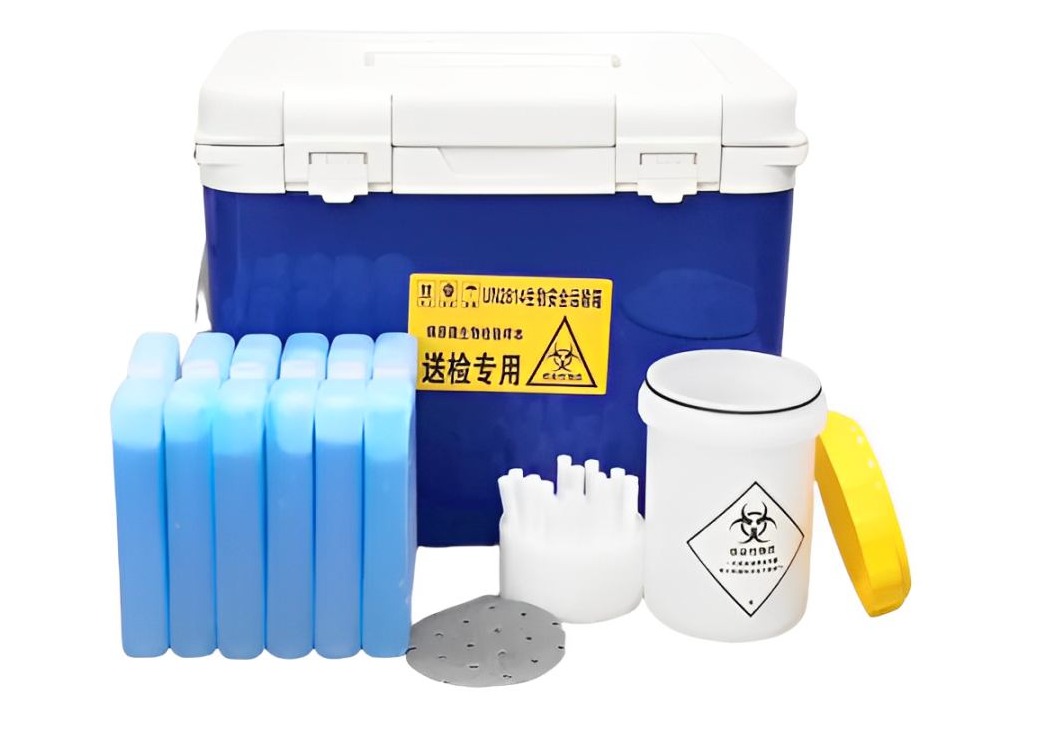
With the FDA framing sample transport as a “national security” issue, customs and health authorities worldwide are stepping up inspections of cross-border shipments. Any sample bag that fails to meet international standards (like UN Regulation PI650 or UN3373 for biohazard materials) risks being held or rejected—delaying trials and wasting valuable specimens.
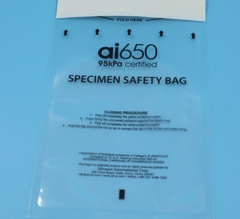
For example, a U.S. biotech recently saw a batch of blood samples detained at a European port because their transport bags lacked 95kPa pressure certification, a key benchmark for leak resistance.
-
Supply Chain Diversification Becomes Critical
The 30% reliance on China’s sequencing services has forced U.S. firms to seek alternative partners in regions like Southeast Asia or Europe. This shift means longer transport routes—and a greater need for durable, temperature-stable packaging that protects samples from turbulence, temperature fluctuations, or accidental drops.
AIC’s clients in Singapore and Germany, for instance, now use our ai650 series for 72-hour transcontinental shipments, with zero leakage incidents.
-
Risk Mitigation Is Non-Negotiable
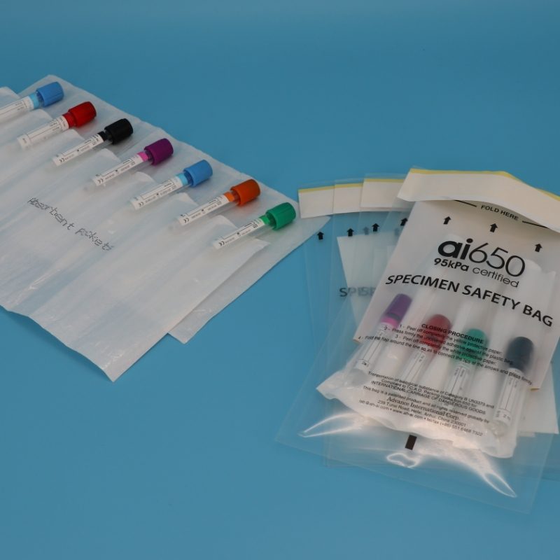
The FDA’s lack of specific “violation examples” has left companies guessing about what qualifies as “high-risk” transport. To avoid gaps, labs and CROs are doubling down on safety features: absorbent pouches to contain spills, tamper-proof seals to prevent contamination, and independent document pockets to keep customs forms separate from samples. These aren’t just “nice-to-haves”—they’re essential for passing audits and maintaining trust with partners.
How AIC’s Solutions Address the FDA Ban’s Challenges
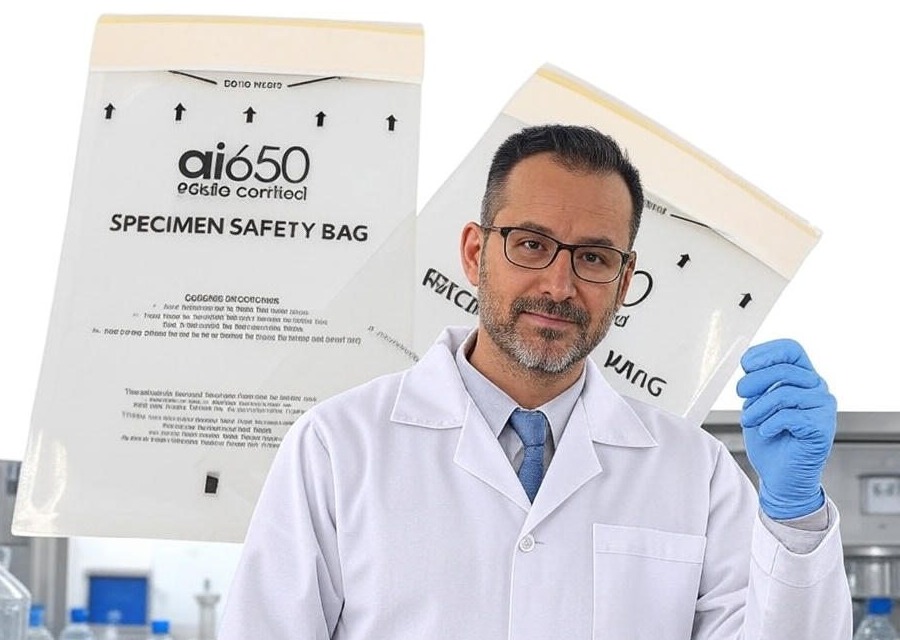
AIC’s product line—built around the 95kPa-certified ai650 series and UN3373-compliant accessories—directly solves the pain points of post-FDA-ban transport. Here’s how we support your operations:
1. UN-Certified Compliance: Pass Inspections Every Time
All AIC specimen transport bags (including the best-selling ai650 model L300420) are factory-tested to meet UN Regulation PI650 and UN3373 standards—the global gold standard for biohazard sample transport. Our 95kPa pressure certification ensures bags resist the physical stress of air cargo (turbulence, pressure changes) and accidental handling, eliminating the risk of leaks that could get shipments detained.
For U.S. firms shipping to alternative partners in Europe: Our bags align with EU 《Animal By-Product Regulation》 (EC 1069/2009), a common requirement for clinical sample imports.
For Chinese labs working with non-U.S. clients: Our compliance with IATA’s Dangerous Goods Regulations (DGR) ensures smooth clearance in 180+ countries.
2. Customized Kits for Diversified Supply Chains
The FDA ban has pushed companies to rethink their transport routes—and one-size-fits-all packaging no longer works. AIC’s customized kit assembly service lets you tailor solutions to your specific needs:
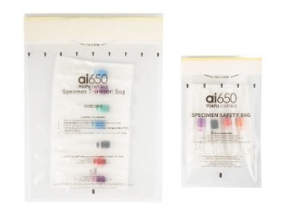
Small-batch shipments (e.g., 6*9inch blood sample bags for regional labs in Southeast Asia).
Large-volume trials (e.g., 7-slotted absorbent pouches (APS4X320) for UN 2814/2900 substances, used by our client Fortrea for cell therapy samples).
End-to-end logistics support: We leverage our Shanghai port proximity (3 hours by train from our Yangtze River Delta factory) to cut delivery times for global shipments—critical for time-sensitive clinical samples.
3. Trusted by Industry Leaders (Even Amid Uncertainty)
In times of policy chaos, partnering with a reliable supplier matters. AIC’s long-standing relationships with top CROs and biotechs—including Parexel, Labcorp, and ERGOMED—speak to our consistency. For example:
A U.S.-based biotech recently switched to AIC’s ai650 bags to ship samples to a new partner in South Korea. Their first 10 batches cleared customs in 48 hours, with no compliance issues.
A Chinese diagnostic lab used our absorbent pouches to safely transport COVID-19 test samples to Australia—meeting both WHO’s biohazard guidelines and local customs requirements.
Beyond the Ban: Building Long-Term Transport Resilience
The FDA’s 2025 policy isn’t just a temporary disruption—it’s a signal that global sample transport will only grow more complex. To stay ahead, biotech firms need partners who don’t just sell packaging, but offer end-to-end peace of mind:
AIC’s R&D team updates products quarterly to reflect new regulations (e.g., recent tweaks to our ai650 seal design to meet FDA’s latest “tamper-evident” guidelines).
Our after-sales team provides 24/7 support for customs questions—critical when shipping to new regions.
We offer OEM/ODM services to help you build branded, compliant kits that align with your global branding (e.g., custom document pockets with your lab’s logo).
Whether you’re a U.S. biotech diversifying your sequencing partners, a Chinese lab expanding to non-U.S. markets, or a CRO managing cross-border trials, AIC’s 95kPa-certified solutions keep your samples safe, compliant, and on time—no matter what policies come next.
Ready to future-proof your clinical sample transport? Contact our team today to design a customized kit for your next shipment. We ship to 50+ countries, with fast lead times and full compliance documentation included.