{ big screen vein finder }

  • The clinical application of venous imaging equipment in oncology departments and chemotherapy wards

    The main value of the venous imaging device in oncology departments and chemotherapy wards lies in addressing the “difficult needle insertion” problem faced by chemotherapy patients due to repeated punctures and deteriorating vascular conditions. It not only significantly improves the success rate of the first puncture, shortens the operation time, but also reduces the pain experience of patients during the puncture process and enhances their satisfaction with medical treatment.

    Clinical research data have demonstrated its specific effects:


    Improving the success rate of one-time puncture: When using an infrared imaging device on elderly cancer patients, the success rate of one-time puncture can increase from 73.6% in the control group to 92.5%. Recent randomized controlled trials also show that using a near-infrared light imaging device results in significantly higher scores for the visibility of blood vessels reported by nurses.


    Shortening the operation time: The use of the imaging device significantly reduces the total time required to locate the blood vessels and successfully perform the puncture. Studies have shown that the puncture time in the group using the equipment is significantly faster than that in the manual group.


    Enhancing patient and nurse satisfaction: Successful punctures directly reduce patients' pain and anxiety. Research has confirmed that patients using the imaging device have higher satisfaction with the nursing procedures, and the puncture confidence of the nurses has also significantly increased.

    These advantages mainly stem from the specific clinical needs of cancer patients:

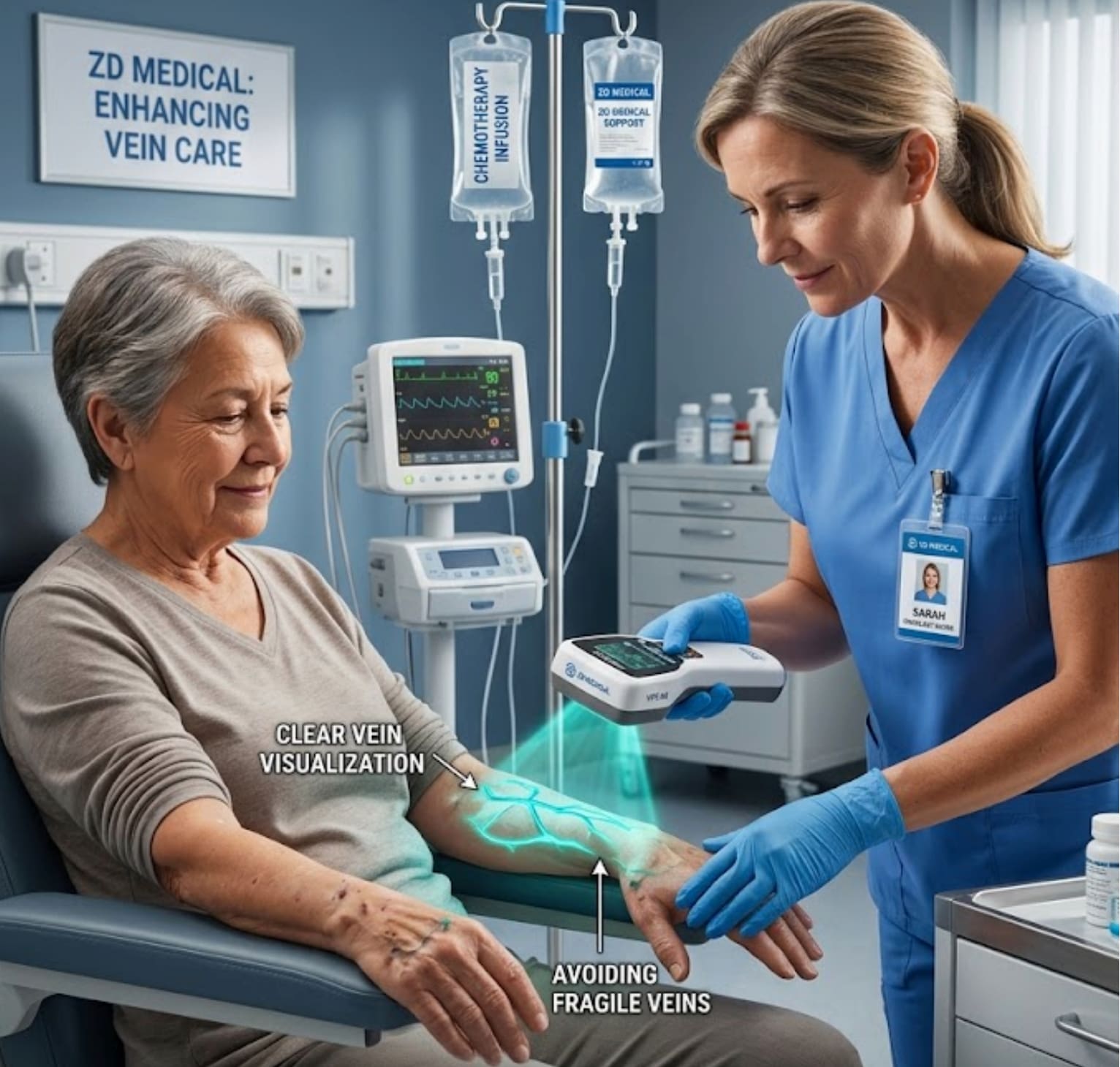
    "Chemotherapy-induced phlebitis" and vascular sclerosis: Repeated damage to the vascular endothelium caused by long-term infusion of chemotherapy drugs leads to hardening, brittleness, and collapse of the veins, making the traditional puncture method relying on vision and touch extremely difficult. The imaging device can penetrate the skin and clearly display the direction and bifurcations of the blood vessels, helping nurses avoid unhealthy vascular segments.


    Protecting limited venous resources: Chemotherapy patients usually require long-term treatment, and every available vein is extremely precious. Using the imaging device to reduce the number of puncture failures can maximize the avoidance of vein damage and extend the lifespan of the patient's venous vessels, preserving the access for subsequent treatments.
    Applicable to elderly and special patients: Elderly cancer patients have loose skin, reduced subcutaneous fat, poor vascular elasticity, and prone to sliding. Studies have specifically confirmed that the infrared venous imaging device has a significant effect in elderly cancer patients, effectively solving these problems.

    Expansion of Applications and Limitations

    Apart from the conventional peripheral venous puncture, the venous imaging technology can also assist in more complex surgical procedures. For instance, a study utilized the dynamic ultrasound imaging technique to perform PICC (peripherally inserted central catheter) catheterization for cancer patients in the ICU, and was able to provide real-time guidance for addressing catheter malposition issues.


    Interested in becoming a distributor? Contact us for wholesale pricing.

    Also welcome to contact us, we are ZD Medical Inc.
    Tel : +86-187 9586 9515
    Email : sales@zd-med.com
    Whatsapp/Mobile : +86-187 9586 9515


  • What are the differences among some different vein finder?

    The following differences also need to be considered when making a choice:
    1. Display mode


    Direct vision/projection: Project the vascular image directly onto the patient’s skin. The advantage is that the operator does not need to shift their gaze and their hands and eyes are in perfect coordination. The drawback is that the projection may deform due to the curved surface of the skin.

    Screen display type: View vascular images on an independent display screen. The advantage is that the image is clear and stable, and can be viewed by multiple people. The drawback is that the operator needs to switch the line of sight between the patient’s skin and the screen.


    2. Portability and form

    Handheld portable: The most common, it looks like a large flashlight or probe, is flexible and convenient, and can be used beside the bed.

    Desktop/floor-standing: Usually more powerful in function, not easy to move, and often used in fixed departments.

    Head-mounted/wearable: Free up hands, but still in the development stage and not widely used.


    3. Functional differences

    Simple positioning vs. navigation guidance

    Positioning type: Only displays the location and direction of blood vessels. Most infrared devices fall into this category.

    Navigation-guided type: It can not only display blood vessels but also provide real-time guidance on the insertion path and depth of the puncture needle through technologies such as laser indication and puncture needle tracking. Most high-end ultrasonic devices are equipped with this function.

    Vascular recognition algorithm: Smart devices can automatically identify and outline the center line of blood vessels through algorithms, and even assess the suitability of blood vessels, rather than merely displaying an image.


    4. Cost and Maintenance

    Near-infrared equipment: The purchase cost is relatively low, almost no consumables are required, and maintenance is simple.

    Ultrasonic equipment: The purchase cost is high, it requires consumables such as coupling agents, and the maintenance and professional training costs are also high.


    How to choose?

    For routine clinical needs (such as outpatient infusion rooms) : High-quality near-infrared light vein finder are the most cost-effective choice and can solve the puncture problems of most patients.


    For patients with difficult puncture (such as ICU patients, the elderly, those undergoing chemotherapy for tumors, and obese patients) : Portable ultrasound vein finder are more effective tools because they can “see” deep blood vessels and have a higher success rate.


    For the establishment of medium to long-term vascular access: Ultrasound equipment is the gold standard, and even an essential tool, as it can assess vascular conditions, prevent accidental arterial puncture, and guide catheter placement in real time.


    For institutions with limited budgets but still hoping to enhance the efficiency of punctures: They can start with mid-to-high-end near-infrared projection equipment, which can intuitively help nurses quickly locate.


    Interested in becoming a distributor? Contact us for wholesale pricing.

    Also welcome to contact us, we are ZD Medical Inc.
    Tel : +86-187 9586 9515
    Email : sales@zd-med.com
    Whatsapp/Mobile : +86-187 9586 9515