OCT shows you structure. OCTA shows you perfusion.
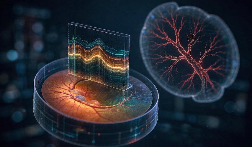
A standard optical coherence tomography (OCT) scan answers the question "what does this tissue look like?" — it renders the retinal layers as a cross-section, micrometer by micrometer. OCT angiography (OCTA) answers a different question: "is blood actually reaching this tissue, and in which layer?" It builds that map from repeated OCT scans of the same location, using the movement of red blood cells as its own intrinsic contrast agent.
Because it needs no dye and no injection, OCTA turns a vascular question that once required a fluorescein angiogram into a two-minute, dye-free, repeatable scan. Nothing structural is replaced. A new dimension is added.
In one line: if structural OCT is a topographic map, OCTA is a live traffic overlay on top of it.
Why the blood-flow layer matters more than it used to
Many of the sight-threatening diseases ophthalmologists manage every day are fundamentally vascular in origin. They begin as a change in perfusion — long before that change becomes visible as structural damage or as something a patient can feel.
|
Disease |
What structural OCT shows |
What the blood-flow layer adds |
|
Diabetic retinopathy |
Retinal thickening once oedema has developed |
Non-perfusion zones and microaneurysms that can precede visible haemorrhage |
|
Wet AMD |
Fluid, intraretinal cysts, subretinal hyperreflective material |
The neovascular membrane itself — its location, shape and extent for treatment planning |
|
Glaucoma |
Retinal nerve fibre layer and ganglion cell complex thinning |
Vessel-density reduction around the optic disc and macula — a functional-supply angle on risk |
|
Retinal vein occlusion |
Cystoid macular oedema |
The exact territory of ischaemia, which drives the laser-vs-injection decision |
|
High myopia / vascular pathology |
Chorioretinal atrophy, layer disruption |
Choriocapillaris flow, as an early marker of progressive myopic damage |
The pattern is consistent. Structure tells you what has already been lost. Perfusion tells you what is under threat now — and that is the window where intervention still changes the outcome.
What OCTA is actually measuring
OCTA does not photograph blood vessels. It detects motion, and infers flow from it.
- The device acquires several OCT B-scans at the same retinal location, milliseconds apart.
- Software compares each pixel across the repeats and flags pixels where the signal decorrelates — the optical signature of moving blood cells.
- Those motion signals are stacked into a depth-resolved volume, then projected into en-face slabs.
- Layer segmentation separates the superficial capillary plexus, the deep capillary plexus, and the choriocapillaris.
Two practical consequences follow from this method, and both matter clinically:
- Flow-dependent. A vessel that is sclerosed, thrombosed or collapsed may carry no detectable signal and will appear absent. The device is not missing it — it is correctly reporting that it is not perfused.
- Static, not dynamic. OCTA produces a snapshot of the vascular network. Unlike fluorescein angiography, it cannot show leakage or the direction and speed of flow over time.
This is why OCTA supplements traditional angiography rather than replacing it outright: OCTA answers where is the vessel and is it flowing, FFA answers is it leaking, and in what sequence.
Where the blood-flow layer changes the decision, not just the picture
Diabetic retinopathy: moving the intervention window earlier
The clinical value of vessel-density mapping in diabetes is not cosmetic. Non-perfusion and capillary loss are the terrain on which proliferative disease develops. Seeing that terrain — and quantifying it — lets a clinician stratify follow-up intervals by actual ischaemic burden rather than by referring to a staging label alone.
Wet AMD: planning the injection before it is given
For anti-VEGF therapy, the neovascular membrane's morphology, its position relative to the fovea, and its response to the previous injection all carry information. OCTA brings those into one co-registered view with the structural scan, so the plan and the monitoring use the same coordinate system.
Glaucoma: a flow-based risk signal
Recent work has strengthened the case for macular perfusion as a non-invasive biomarker in glaucoma. One 2025 cross-sectional study of 114 participants. Superficial plexus metrics showed a moderate but significant association with focal loss (R² = 0.51).
The takeaway for a clinical reader is measured but real: perfusion metrics can sit alongside structure as a second, complementary axis — useful for early detection and for monitoring, not a stand-alone diagnostic.
How the technology attaches to clinical workflow
On ZD Medical's Luminor OCT platform family, the workflow question is deliberately framed around throughput. A clinic does not need more images; it needs images that can be interpreted and reported inside the visit.
Luminor-D60 is the platform positioned for broader clinical use — higher image clarity, smoother scan capture, and analysis software aimed at identifying retinal disease early enough to reduce missed diagnoses.
Luminor-A80 and Luminor-D60M are directed at outpatient fundus screening and the routine OCTA examination-and-analysis workflow: straightforward operation, clear imaging, and vendor analysis software covering common clinical needs.
The design goal across the family is the same three-part lens that every diagnostic purchase should be judged against:
- Does it capture fast enough to be routine? Scan speed and eye-tracking determine whether the exam fits a busy clinic list or becomes a special appointment.
- Does it repeat well? Reproducibility is what makes serial comparison meaningful; a beautiful single image that cannot be repeated is a discovery, not a monitoring tool.
- Does it produce a report, or just an image? Automated, reproducible reporting is where the efficiency argument is actually won.
How to evaluate the blood-flow layer before you buy
Not every platform that offers an "OCTA mode" delivers usable perfusion data at 9 a.m. on a full list. Ask for these seven things during evaluation:
- Motion-artefact rejection. Test on a patient who cannot hold fixation. If the software cannot suppress saccade and blink artefacts, everything downstream is noise.
- Segmentation accuracy. Verify the superficial and deep plexus boundaries against a known case. Mis-segmentation silently corrupts density values.
- Projection-artefact handling. A large vessel above a capillary slab casts a shadow. Confirm the platform corrects for it, or density will read falsely low.
- Co-registration with structural OCT. Confirm perfusion and structure share one coordinate system across visits.
- Quantification, not just display. Vessel density, flow index, and non-perfusion area should be output as numbers that can be tracked over time.
- Repeat-scan coefficient of variation. The single most important number for monitoring use — and rarely offered in brochures.
- Certification and service for your market. Verify CE / NMPA status, warranty terms, and spare-parts and training support before purchase.
Common mistakes that mute the benefit
Using OCTA as a screening photo. Its strength is depth-resolved perfusion, not a widefield snapshot. Use it on the question it was built to answer.
Comparing across vendors without a repeatability figure. Density numbers are not interchangeable between devices. Trend within one platform, not across several.
Skipping artefact training. Most "abnormal" perfusion scans that turn out to be normal are artefact, not pathology. Budget time for clinical onboarding — it changes image quality more than any hardware upgrade.
Treating it as a replacement for FFA. Leakage, and dynamic flow, remain FFA's questions. OCTA is complementary, and should be positioned as upstream and more frequent.
Frequently asked questions
Is OCTA safe? OCTA uses near-infrared light and requires no dye and no injection. It is non-invasive and non-ionizing, and because it needs no contrast agent, it avoids the systemic and allergic risks associated with fluorescein angiography.
Can OCTA replace fluorescein angiography? No — they answer different questions. OCTA maps perfused vessels in depth; FFA shows leakage and temporal flow. In most clinical pathways the two are complementary, with OCTA used more frequently because it is repeatable.
Do patients need dilation for an OCTA scan? It depends on the device and the imaging target. Many macular scans can be acquired without dilation, but image quality and signal strength determine the artefact burden — a practical point to confirm with each vendor during evaluation.
How long does an OCTA scan take? Acquisition is typically measured in seconds per volume, which is what makes it feasible to perform within a routine outpatient visit. Total chair time depends on capture repeatability.
Is OCTA useful in glaucoma? Increasingly, yes — as a complementary metric. Vessel-density studies, including the 2025 data cited above, support macular perfusion as a non-invasive biomarker for early detection and monitoring alongside structural OCT.
What is the difference between superficial and deep capillary plexus?
The superficial capillary plexus sits in the ganglion cell layer; the deep plexus lies in the inner nuclear layer. Both are segmented from the same OCTA volume. Because they can be affected differently by disease, reporting them separately matters for both detection and monitoring.
Where this leaves the clinical argument
The blood-flow layer is not an upgrade, a mode, or a marketing feature. It is a different measurement domain. Structure and perfusion together describe both what a tissue is and whether it is being supplied — and it is the second half of that sentence that usually determines whether vision is preserved.
For clinics building a modern imaging pathway, the practical sequence is straightforward: establish a repeatable structural baseline, add depth-resolved perfusion on the same coordinate system, and quantify both so change over time is visible rather than inferred.
Key takeaways
- Structural OCT = anatomy; OCTA = perfusion. They answer different questions and belong together.
- The blood-flow layer often changes first, which is why it can move the intervention window earlier.
- Method determines limits: flow-dependent (so non-perfused vessels appear absent) and static (so leakage is still FFA's question).
- Quantification and repeatability decide clinical value — not image beauty.
- Evaluate artefact handling, segmentation and co-registration before buying, not after.
About ZD Medical
ZD Medical Inc. develops and manufactures ophthalmic diagnostic imaging systems, with R&D centres in Los Angeles (optical systems) and Silicon Valley (OCT image research and processing). The Luminor OCT/OCTA platform family is engineered for outpatient screening, retinal disease identification and routine clinical analysis. Vein visualization and dry-eye diagnostic lines complete the portfolio.
Interested in becoming a distributor? Contact us for wholesale pricing.
Tel / WhatsApp: +86-187 9586 9515 · Email: sales@zd-med.com